
Achieve Specialized Precision with the New SPECT/CT Viewer
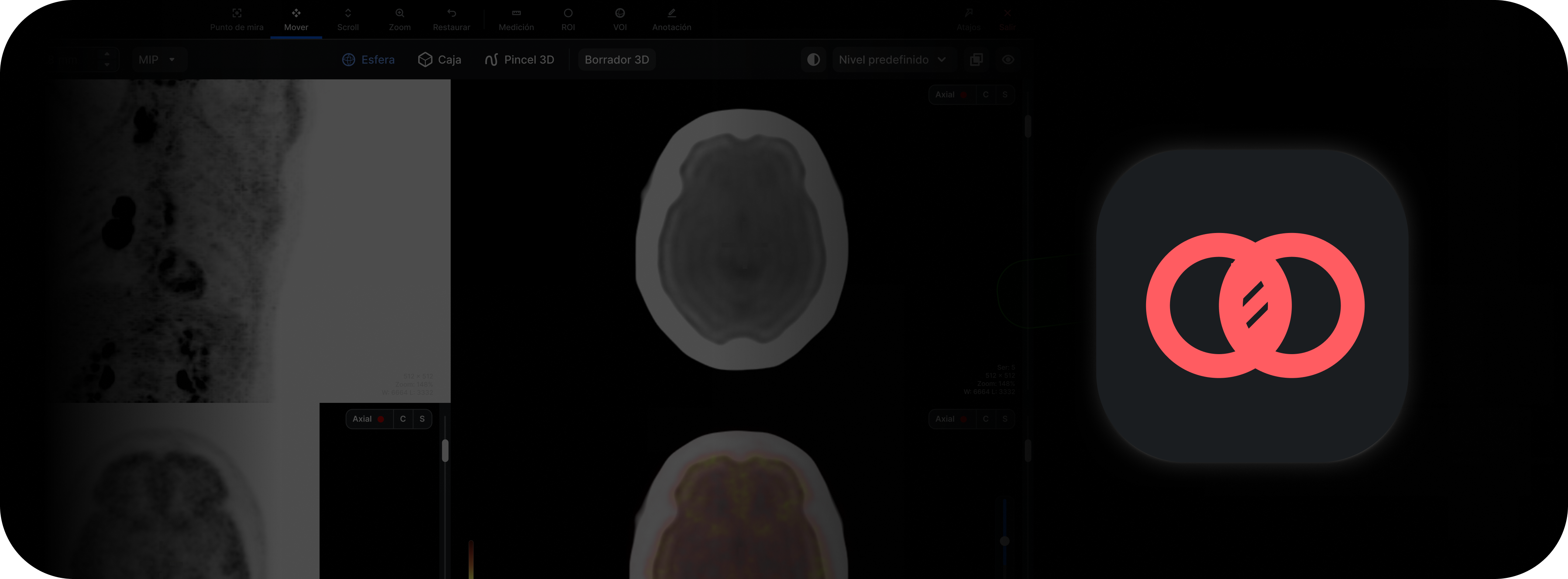
As part of the enhancement of the viewer's advanced capabilities, it is now possible to perform SPECT/CT image fusion directly within the RIS-PACS system. This feature extends previous capabilities, which were limited exclusively to PET/CT fusion, and broadens the viewer's clinical utility in nuclear medicine and oncology.
What it is
This is a tool that allows the radiologist to overlay functional images obtained by SPECT with the anatomical images from a CT, achieving an integrated visualization. The system leverages the spatial registration information included in the DICOM series to automatically align both modalities and display the study jointly in the viewer.
What it is for
SPECT/CT fusion allows:
- Correlating functional findings with their precise anatomical location.
- Improving interpretation in oncology, cardiology, and neurology.
- Performing faster, more reliable evaluations without the need for external software.
- Ensuring that the radiology report includes both metabolic and structural information.
How it affects the workflow
Previously, users had to use specialized workstations or additional software to fuse SPECT with CT. Now, the integration can be performed directly from the RIS-PACS viewer, within the same reading workflow, without switching environments or losing continuity. This reduces interpretation times and avoids dependence on external workstations.
What changes compared to previous versions
Until now, fusion was only available for PET/CT. With this update, it is also enabled for SPECT/CT, provided that the series sent from the equipment include the necessary spatial information. In studies where this is not met, the viewer will display a warning or will not allow the overlay.
Implications for the user
In SPECT/CT studies, it is important to keep in mind that nuclear medicine equipment generates different reconstruction series: an "uncorrected" one (without attenuation map) and a "corrected" one (Corrected/AC), which is the one aligned with the CT. Depending on the manufacturer, not all series include the spatial data required for automatic fusion. For this reason, it is always recommended to review and load the attenuation corrected or SPECT/CT Registered series when available, since it is the only one that can be displayed correctly fused with the CT.
Final considerations
The fusion of SPECT/CT studies expands the clinical capabilities of the RIS-PACS viewer and improves the integration of functional and anatomical information in nuclear medicine settings. Its correct use depends on properly selecting the series sent and on the configuration of the acquisition equipment.
For optimal performance, it is recommended to coordinate with the nuclear medicine department to standardize series export and ensure the availability of the "attenuation corrected" series.