Clinical, Cultural, Computational, and Regulatory Considerations to Deploy AI in Radiology: Perspectives of RSNA and MICCAI Experts
Summary
The Radiological Society of North of America (RSNA) and the Medical Image Computing and Computer Assisted Intervention (MICCAI) Society have led a series of joint panels and seminars focused on the present impact and future directions of artificial intelligence (AI) in radiology. These conversations have collected viewpoints from multidisciplinary experts in radiology, medical imaging, and machine learning on the current clinical penetration of AI technology in radiology and how it is impacted by trust, reproducibility, explainability, and accountability. The collective points—both practical and philosophical—define the cultural changes for radiologists and AI scientists working together and describe the challenges ahead for AI technologies to meet broad approval. This article presents the perspectives of experts from MICCAI and RSNA on the clinical, cultural, computational, and regulatory considerations—coupled with recommended reading materials—essential to adopt AI technology successfully in radiology and, more generally, in clinical practice. The report emphasizes the importance of collaboration to improve clinical deployment, highlights the need to integrate clinical and medical imaging data, and introduces strategies to ensure smooth and incentivized integration. Keywords: Adults and Pediatrics, Computer Applications–General (Informatics), Diagnosis, Prognosis © RSNA, 2024
Key points
- Artificial intelligence (AI) tools can play a key role in radiology if radiologists trust in their design, deploy them with adequate training, and establish clear guidelines regarding clinical accountability.
- For successful AI deployment, radiologists and AI scientists should define a unified agenda, language, and set of expectations.
- Clinical institutions should align their staffing, data flow, and computational resources to deploy and monitor AI systems.
Introduction
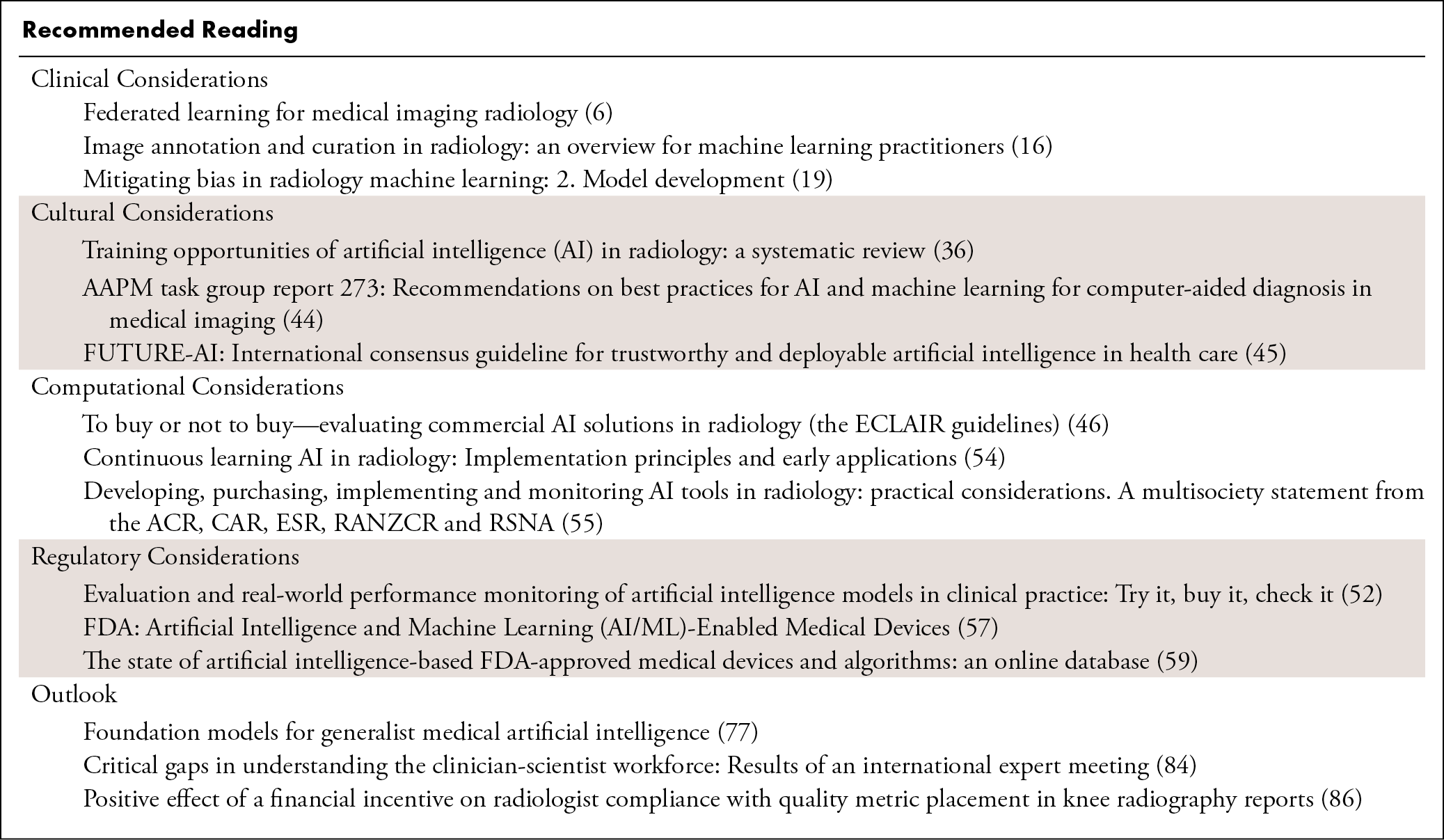
This special report is the third in a series of articles based on joint panels and seminars by members of the Radiological Society of North America (RSNA) and the Medical Image Computing and Computer Assisted Intervention (MICCAI) Society that focus on the present impact and future directions of artificial intelligence (AI) in radiology 1,2. The current report addresses the clinical deployment of AI into the practice of diagnostic radiology and explores the clinical, cultural, computational, and regulatory considerations that imaging experts, clinical and technical, should consider for successful adoption of AI technology. Recommended reading relevant to each section is provided in the Table in the article. This report also introduces strategies to ensure smooth and incentivized integration of AI into the radiology workflow.
Clinical Considerations
Tools to exchange data either for centralized analytics or distributed learning will be able to support sharing of data and/or models but will require additional institutional infrastructure and support.
Image annotation may become a less substantial hurdle in the next 5 to 10 years.
AI models built with small datasets or in-house data can navigate challenges related to data bias and diversity if they are applied to targeted populations and carefully monitored.
Data Sharing and Analytics
Data sharing is crucial to foster the development of machine learning (ML) models. Many institutions are not allowed or are unwilling to share their data, and there is a critical shortage of data on rare diseases 3. Clinicians may feel uncomfortable sharing images because of lack of time and expertise to extract, anonymize, and upload large volumes of data efficiently. Thus, data sharing can limit the deployment of AI into clinical practice and can pose a substantial barrier between institutions and even within the same institution. Data sharing also must abide by ethical and legal standards 4,5.
When institutions are open to data sharing, methods for data collection and transfer must be secure. Email is insecure and inefficient, compact discs are time-intensive and require physical mailing, and hard drives require exchange of physical media. File-sharing platforms are more secure than email but can be slow and require multi-institutional information technology (IT) approval for external collaborations. A centralized research picture archiving and communication system (PACS) can be expensive and can have a limited array of data-analytic tools. Open-source and commercial platforms are available that incorporate AI, but tools are still developing. Cloud-based servers with AI capabilities require expertise to make sure data are secure and Health Insurance Portability and Accountability Act compliant. For institutions that are not open to data sharing, federated learning can facilitate AI model development and deployment without data exchange 6–8, but federated learning facilities are not commonly available in radiology departments and require specialized expertise 9.
Data Annotation
AI relies on well-curated and annotated data. Annotation is time-consuming and labor-intensive and poses challenges for large datasets 10. Multiple annotators are required to tackle large datasets and allow measurement of interrater variability. The RSNA AI challenges have shown that expert annotators can exhibit substantial variability even when the annotation tasks are well-defined, highly documented, and carefully communicated 11–14. Thus, it is important to record and make available all details of the annotation process, including methods for adjudication and quality assurance, to those who are evaluating AI tools for clinical deployment.
Annotation of radiologic images often requires transferring images to a research computer that has annotation software, potentially requiring uploads, and only then starting the segmentation or other type of annotation. Some institutions have been able to work with their IT departments to directly transfer data from the clinical PACS to the annotation software 15, but most do not have such capabilities. Thus, a major hurdle for annotation is the lack of institutional infrastructure for data access and sharing with annotators. The availability of web-based, easy-to-use annotation tools allows radiologists from around the world to contribute their expertise and is key to reducing hurdles in annotations once the de-identified data can be secured from the contributing institutions.
On the other hand, there has been substantial evolution in our understanding of annotation and the level of quality required for AI development 16. Although very high-quality testing data are important, noise can be tolerated in the training data, providing it does not contain bias. New technologies for active learning, iterative annotation, and self-supervised learning enable AI models to learn more rapidly from fewer annotations.
Bias and Model Performance
ML models often learn biases in the training data. Underrepresented characteristics, such as image acquisition features or patient features, can be especially prone to bias. Potential sources of bias differ for various AI applications and depend on the clinical setting in which the AI tool will be used. Diversity of background and experience in both the development and deployment teams is helpful to identify and address biases. Because it is not feasible for an AI model to work everywhere reliably, it is important to recognize the intended use and potential limitations of the model 17–19. For instance, an institution may want to develop a model for in-house use only. In that case, it will be practical to develop a model that performs well only on internal data. However, the bias and limitation of the model, likely not applicable to underrepresented populations in research, must be clearly disclosed. It is critical to continuously monitor performance, identify changes in performance, and provide detailed and updated documentation.
Cultural Considerations
Training radiologists to work with AI tools will become routine.
Motivation for radiologists to use AI is key to increasing clinical efficiency and to allowing them to perform complex tasks, such as measuring volumes, predicting outcomes, and integrating imaging data with the broader electronic health record (EHR).
AI tools must be designed with the trust of radiologists and clear definitions of clinical accountability.
Volumetrics
Automation and AI in radiology potentially can provide routine three-dimensional (3D) volume measurements, which represents a cultural shift from two-dimensional (2D) measurements such as the Response Evaluation Criteria in Solid Tumors (ie, RECIST) 20–22. For most radiologists, qualitative image interpretation and 2D measurements are still the standard of care in clinical practice and in radiology reports. Studies have shown the benefits of volumetric measurements, such as in cancer imaging, where 3D volumetrics offer better outcome prediction and treatment response evaluation than qualitative assessment or 2D measurements 23–27. Without AI, such measurements require a substantial amount of additional time. To prepare radiologists for volumetric data interpretation, radiology training should emphasize a shift to 3D from 2D measurement, and dedicated education on lesion contouring should be incorporated into radiology training.
Many radiologists and other specialists would like to have better 3D measurement tools. However, few 3D tools are available at every radiologist’s desktop, although there is an increased availability of contouring tools in clinical PACS. In addition to contouring tools, there is a critical need for tools that allow individual lesion tracking, for example, in the setting of metastatic disease. Thus, there is increasing potential for volumetric tools—powered by AI and easily adjustable by radiologists—to play a key role in clinical care.
Image Evaluation to Outcome Prediction
Outcome prediction is a valuable end goal. AI-based prediction of cancer mortality, cardiovascular morbidity, or organ function, among others, can be competitive with routinely available clinical methods 28–30. Outcome prediction also may motivate radiologists to focus on data interpretation with the help of AI. To be clinically relevant, outcome prediction needs to be performed in a clinical scenario where it has the potential to change care. There are risks in outcome prediction, too. Models may be biased in ways that human experts cannot appreciate easily 31. If the task is image evaluation, it is more likely that a human reader would catch the error. But if the model is expected to have superhuman performance (say 5-year risk prediction for a task that a human cannot do), the model deployment must be carefully monitored to alleviate risks.
Human-Machine Interaction
In daily practice, radiologists’ measurements can exhibit substantial intraobserver and interobserver variability 32–34. AI-based algorithms may reduce measurement variability and may allow greater consistency. However, automation bias can increase error rates in the setting of AI utilization, and human-AI interactions are understudied. Even if an AI model works well, there is no guarantee radiologists will use it correctly to improve clinical performance. Human-AI interactions and their cultural and clinical implications require further research to ensure optimal performance 35. The design of stronger and more comprehensive programs to train radiologists to work with AI approaches is important, and these programs should be integrated into early career curriculum 36–38.
Clinical Integration
Radiology AI systems can be integrated with currently used software to reduce the effort of interpretation and reporting 39. Ideally, AI should reduce the effort of interpretation and reporting. A major characteristic of radiologists’ daily workflow is the long list of studies that need to be read every day, which makes the radiologist prioritize efficiency in reading studies. Any activities outside of reading can be a substantial distraction.
AI tools within PACS provide an efficient method for radiologists to adapt them into clinical practice. Integration of AI into PACS allows manual corrections of segmentations that would not be possible otherwise, thus providing another level of safety checks. AI tools can prioritize worklists: They can identify studies with acute findings or automatically route studies with specific pathology to the best-matched specialist radiologists 40.
An important challenge in radiology is the limited clinical information provided when a study is being interpreted. Although an EHR is available, its organization may not allow one to extract information easily. Therefore, integration of EHRs with AI tools in radiology has great potential for clinical practice 41. AI tools that extract and summarize clinically relevant information from the EHR can support more precise interpretation of studies and improve radiology reports.
Trust and Accountability
Trust is important in clinical practice. To assure patient safety, the field of medicine is more conservative than others in adopting new technology 42. Moreover, AI applications are not always intuitive, and “black box” models that lack explainability are difficult to trust. As such, few clinicians trust AI for autonomous evaluation, and AI deployment strategies must incorporate radiologist supervision for the near future. Radiologists are, for the most part, comfortable to be held accountable for studies they interpret with or without AI support 43. For example, if the AI tool highlights a positive finding, the radiologist can visualize and confirm the finding with confidence. Exceptions to explainability should be considered, such as for identification of radiomics features that cannot be perceived by the human eye.
An integrated approach to build trust should include retrospective validation with local data (ie, test on your own data to trust it) and prospective evaluation using AI in practice. The latter is tied to having high positive and negative predictive values. Because algorithms are not error-proof, it is important for radiologists to understand how errors may happen and that they are something to be expected. To earn the trust of clinicians, AI algorithms also must support the clinical workflow (eg, automatic measurements incorporated into radiology reports) and expedite the turnaround of studies. AI methods where model predictions are not repeatable, do not generalize within the scope of their development, or are not well calibrated should not be implemented. These and other concerns on accountability, quality control, and trust have been addressed in publications by leading experts from international societies including RSNA, MICCAI, and the American Association of Physicists in Medicine 44,45.
Computational Considerations
Cloud computing may be most effective for radiology departments that lack hardware and maintenance resources.
The design, development, deployment, and monitoring of radiology AI tools should be done by data scientists and radiologists working together.
Clinical institutions must prioritize the data flow from acquisition devices to PACS and/or data lakes and to AI servers.
Hardware Availability
To deploy AI in clinical settings requires consideration of computational factors for training and inference, such as hardware availability 46. In most clinical centers, and especially in limited-resource settings, the cost and maintenance of graphics processing unit servers and high-performance computing resources limits their availability 47. Approaches such as cloud-based systems may be necessary. The use of commercial algorithms in the cloud or homegrown algorithms on-premises can also present challenges. However, if graphics processing unit memory requirements can be reduced for AI models during inference, hardware availability should not be a substantial challenge. Overall, although hardware availability may depend on the location and use case, less expensive central processing units may be sufficient for many AI applications.
AI Scientists in Radiology Departments
Communication between radiologists and AI scientists is key to incorporate AI into clinical practice. Radiologists are highly skilled at characterizing phenotypes and labeling images but often lack the tools to annotate datasets and build databases 48. AI scientists, on the other hand, have skills in image processing and algorithm development, but they often focus on questions that may not have the highest yield in clinical practice. It is crucial to encourage communication between radiologists and AI scientists through grant mechanisms, conferences, and professional and social gatherings to build trust and collaboration.
The shortage of skilled AI scientists in radiology departments poses another limitation. Academic medical centers frequently compete with technology companies for data scientists. To create robust and reliable AI models, it is important to have AI/data scientists, software engineers, PACS/radiology information system specialists, and radiologists working together 2,49.
AI Performance
In general, clinical accuracy is more important than speed 50. If inference time is a substantial problem, one can limit the model’s size to not exceed a given time per inference and train and tune that model to the best performance possible. In many applications, speed is not particularly relevant: A computation time of a few minutes generally will not be noticeable. One exception is for tasks such as interactive segmentation, where the user must wait for the AI system to respond. As algorithms increase in accuracy with the availability of larger training datasets, it will be possible in many scenarios to use less complex AI models. Less complex models will lead to more rapid computation and distribution of AI results to the clinic and the need for a framework to evaluate their performance 51. In many clinical settings, the primary bottleneck is not computational time, but rather the time to transfer data from acquisition device (eg, CT scanner) to PACS to AI inference server.
Radiology practice is fast-paced, dynamic, and often stressful; it requires algorithms that can keep up with the demands of clinical practice. AI algorithms can be incorporated into practice automatically or activated manually by a radiologist 52. Although an algorithm’s accuracy is important, consistency of measurement is its inherent advantage 53. The qualities of an algorithm need to be evaluated in the context of the clinical problem and use case. Domain shift and algorithm generalizability remain challenging questions for research and clinical implementation.
Continuous Evaluation
Continuous evaluation and quality control of AI tools is a critical aspect of clinical implementation 54. Monitoring of performance is not trivial and is currently a weakness for most AI systems. Centralized datasets that can be used as “phantoms” to test algorithms and image processing software may provide one solution. Technologies for continuous evaluation must not require explicit supervision or labels. Therefore, regulatory guidelines should assure continuous evaluation 55. Most organizations lack the staff to perform these evaluations effectively. For models with access to reference standard data, continuous evaluation and retraining could be automated. For models without such data, expensive and time-consuming annotation could be limited by focusing validation on new images in the event of a scanner replacement or protocol change.
Regulatory Considerations
Radiology is the leading application field of U.S. Food and Drug Administration (FDA)–approved medical AI devices.
Vision and language models can positively impact the field of radiology and should undergo regulatory oversight.
The role of regulatory approval in the implementation of AI tools in clinical practice will require ongoing consideration.
Regulatory Guidelines
The field of medical AI is rapidly evolving, and regulatory agencies such as the FDA are striving to keep up with the pace of innovation 56. Notably, the development of large foundation and generative AI models is progressing fast, including vision and language models, which are AI tools that integrate understanding of images and text. Nevertheless, technology development outpaces the creation of regulatory guidelines; as of October 2023, there was no FDA-authorized device that used generative AI 57. As a result, commercial development favors some types of algorithms over others. For example, the computer-aided triage pathway has seen relatively high rates of FDA clearance, which has resulted in a flood of worklist prioritization tools in the market. Similarly, time-limited financial incentives such as new technology add-on payment (ie, NTAP) reimbursement have driven demand for tools in the hope of an early financial windfall that has largely been exaggerated. There is also concern that many FDA-approved algorithms have not undergone sufficient external or prospective validation. Published reports of performance may not accurately reflect how well these algorithms perform in real-world settings 58.
Regulatory Approval
Regulatory authorization is critical for the rollout of AI tools in clinical practice 59. By July 2023, 79% of the medical AI devices authorized in 2023 by the FDA were in radiology, followed by cardiology and neurology 57. There is support for the authorization of software as a medical device, which allows for innovation and does not stifle progress. FDA clearance is not required to use locally developed AI tools; it focuses on regulating marketing claims and commercialization. Moreover, marketing claims and FDA submission materials are not always concordant. However, strong oversight and understanding by practitioners of potential harms, particularly in vulnerable populations, is important. Regulatory approval is needed for potential financial reimbursement for the use of AI and facilitating the transition from research to clinical practice.
Outlook
Large and general foundation and generative AI models, including vision and language models, can impact the clinical adoption of AI tools and may help reduce burnout in radiology.
Financial incentives to use AI will encourage hospitals to invest in AI-based software and increase the motivation for clinicians to use new technologies.
To advance radiology AI tools in clinical care and research, multidisciplinary societies can adopt a unified agenda, language, and set of expectations.
Impact of the COVID-19 Pandemic
The COVID-19 pandemic has had a mixed impact on the clinical adoption of AI 60. On the positive side, the intellectual and clinical challenges of this new, dangerous, and highly contagious disease spurred radiologists and AI scientists around the world to create algorithms for diagnosis and triage 61,62 and to establish new data repositories like the Medical Imaging and Data Resource Center (ie, MIDRC) 63. The increase in imaging volume, coupled with the pre-existing shortage of radiologists, made radiologist burnout a more widespread issue and encouraged radiologists to explore AI tools to increase efficiency and accuracy in clinical practice. The desire to quickly integrate COVID risk models yielded an expedited pathway to move tools from research into the clinical realm. The pandemic also presented barriers to the clinical adoption of AI, as new software could not be implemented easily due to restrictions on travel and physical presence in the hospital. COVID applications have shown how AI models can be brittle and not generalize 64.
Although some regulatory obstacles were removed, at least temporarily, COVID has not impacted the availability of algorithms that solve real clinical problems. Researchers have moved away from simple segmentation toward clinically useful predictions—such as whether a patient will require hospital admission, will require artificial ventilation, or will have a higher risk of death—but clinicians have not adopted the AI assessments into clinical care with the same enthusiasm.
What Are AI’s “Low-hanging Fruit” for Radiology?
AI has a huge potential to transform medical imaging in various ways by integrating imaging and clinical data in relational databases that can be used for both clinical practice and research, not just for billing 65,66. It is important to seamlessly link Digital Imaging and Communications in Medicine (ie, DICOM) data with Fast Healthcare Interoperability Resources (ie, FHIR) data from the EHRs within PACS 67,68. Additionally, data scientists and clinicians need to apply the existing well-validated AI tools, such as organ and lesion volumetrics, to the clinical setting. Examples include liver, spleen, and kidney volumetry, visceral fat assessment, and muscle analysis for sarcopenia and myosteatosis 69. Finally, there is need to personalize the treatment options for each patient with cancer by using AI analytics for cancer radiomics and longitudinal tracking of individual lesions 70,71.
What Is Next in AI for Radiology?
The next frontier for medical imaging AI is implementation in clinical practice 72. Collaboration is essential among radiologists, referring clinicians, data scientists, and health IT professionals to identify and evaluate AI solutions for various clinical scenarios. New metrics would be useful to measure AI’s impact on workflow efficiency, assess clinical outcomes in radiology and related fields, and conduct randomized clinical trials to assess the utility of AI algorithms 55. Promising research directions include unsupervised and self-supervised learning techniques to develop generalizable models that can be adapted to small or niche datasets or to any hospital dataset 73–75. Researchers are also creating broader AI models like foundation models for tasks such as normal versus abnormal detection, rather than narrow supervised ML tasks 76–78. Particularly, large vision language models promise new and general-purpose AI applicability in diagnostic imaging through multimodal data analysis to provide a natural language interface for radiologists 79. Through these developments, AI can play a crucial role in reducing burnout in radiology if the integration and adoption of AI-driven technologies is done through adaptive and user-friendly tools 80.
How Do We Teach and Incentivize Radiologists to Work with AI?
The integration of AI into radiology practice requires educating and motivating radiologists to work with AI 81. AI must be easy to use within familiar software and compatible with the clinical workflow. Radiologists should learn how to evaluate, interpret, and apply AI algorithms for tasks such as segmentation, detection, and classification, as well as how to handle and report imperfect algorithms 82. Radiologists also should learn how to design and conduct clinical trials to assess the impact of AI on clinical outcomes; such training is often lacking in radiology compared with other fields such as oncology. The RSNA’s Clinical Trials Methodology Workshop teaches radiologists about clinical trial design, funding, and implementation. Radiologists also should learn how to communicate and collaborate with AI scientists without feeling intimidated 83. Most radiologists in training are already interested in AI, but they lack a good training mechanism and curriculum. Dedicated training in informatics and involvement in societies such as MICCAI and the Society for Imaging Informatics in Medicine will increase radiologists’ knowledge of AI.
There is also a technical gap between physicians and AI scientists that needs to be bridged 84. One way to facilitate the adoption of AI is to use model cards, which describe an AI model’s purpose, usage, and limitations 85. Another way is to solve real clinical problems, expand the possibilities of data analyses and interpretation, demonstrate value in prospective trials, and improve workflow efficiency, which would make radiologists eager to use AI without needing additional incentives. A third way is for payers to provide financial incentives to use AI, which would encourage hospitals to invest in AI-based software and increase the motivation for radiologists 86.
Unified AI Agenda across Societies and Disciplines
The advancement of AI in medical imaging depends on collaboration across societies and disciplines, which poses both challenges and opportunities. To foster collaboration, it is important to be inclusive, to think beyond personal needs, and to adopt a humanistic and scientific approach to life. Medical imaging societies have collaborated to establish ethical frameworks for AI deployment in radiology and educate clinicians about the potential benefits and pitfalls of AI implementations 55. However, differing agendas, languages, and expectations among societies and medical specialties may hinder communication and limit cooperation 11. Therefore, it is essential to create platforms and mechanisms that facilitate the exchange of ideas and information, such as educational programs, challenges and competitions, and hackathons offered by several societies, including joint initiatives of MICCAI and RSNA. Moreover, it is necessary to educate and motivate both technical and radiology societies to understand the capabilities and limitations of AI, the true clinical needs and problems, and the technical skills and methods required to develop and deploy AI solutions. Finally, it is desirable to pursue innovation and discovery by creating and evaluating novel algorithms, integrating and analyzing imaging and clinical data, and developing and applying AI tools for various tasks and domains. By collaborating across societies and disciplines, AI in medical imaging can achieve its full potential and provide value for clinical practice and research.