
Whitepapers
The latest capabilities of the Diagnostic OS


Browse all
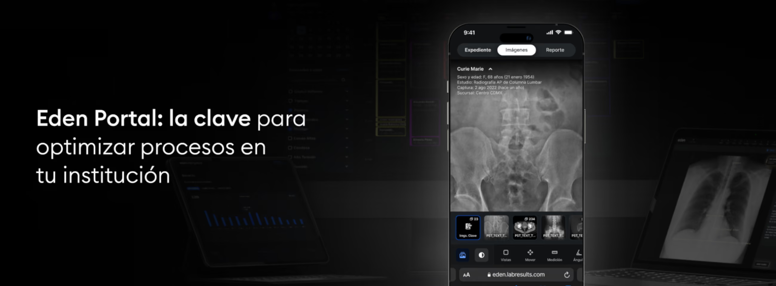
Eden Portal: The Key to Optimizing Processes at Your Institution
Many industry leaders warn that unintuitive medical portals can create confusion among patients and generate more work for staff, disrupting both operational efficiency and the quality of medical care.
Eden Portal is here to change that, acting as a strategic ally that optimizes and simplifies key operations at your institution, from diagnosis to results delivery.
Optimizes results delivery
Eden Portal is a meeting point that connects your institution and your patients, from anywhere, from any electronic device. The ability to send radiology results directly via WhatsApp, securely and quickly, eliminates the need for phone calls, in-person appointments and complex web portals. This not only facilitates continuity of care but also reduces results delivery times by up to 60%.
Reduces the burden on your administrative area
Eden Portal centralizes and improves the storage of clinical information. Now, patients can access their medical history with a tap on their mobile phone.
By consolidating this information on a single platform, it becomes possible to use resources that would otherwise be dedicated to maintaining physical or digital records, enhancing the patient experience and the workflow starting at reception. This also minimizes errors from duplication and data loss, building more reliable processes for your institution.
Streamlines and empowers your radiologists' processes
Eden Portal is not only in the hands of your patients: it is also in the hands of your radiologists. With advanced tools such as 3D reconstruction, vessel analysis and PET-CT on a mobile device, it is possible to perform accurate diagnoses with great agility and address emergency cases instantly.
The power and speed of Eden Portal, even without a direct internet connection, ensure a frictionless workflow, minimizing technical problems and doubling radiologists' productivity.
Eden Portal is not just a patient portal, but a key tool for transforming management at your healthcare institution. Its intuitive interface and 100% mobile viewer make it possible to simplify and optimize every stage of the workflow, from administrative processes to diagnosis and results delivery. With every detail meticulously designed to give your institution a competitive edge, Eden Portal turns what used to be a challenge into an effective solution.
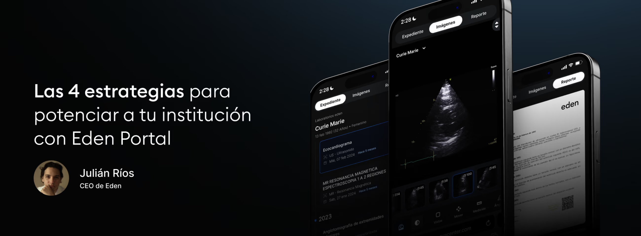
The 4 Strategies to Empower Your Institution with Eden Portal
Today, Eden Portal is raising the standards of our industry in patient and physician relations. With the first 100% mobile DICOM viewer, a complete clinical history at the touch of a finger, and an intuitive interface, this portal transforms the patient experience and places your medical institution at the center of the healthcare universe.
However, every medical technology comes with certain adoption challenges. It is not as simple as pressing a button and saying "now I have Eden Portal": it requires operational changes, system adjustments and, above all, an institutional commitment to implementing new technological tools.
That is why, today, we will share 4 key tips to help your institution maximize the potential of Eden Portal and turn it into the backbone of its growth strategy.
1. Ensure complete traceability of patient information
A key factor in maximizing the use of Eden Portal is ensuring that all patient information, including the unique identifier and full name, is standardized across all systems. This is easily achieved with Eden Management, the advanced cloud-based RIS available to all Eden clients.
Eden Management automatically integrates patient information, from order generation through modality, preventing errors and ensuring that patients can view their complete history in the portal. However, we know that some institutions have specific points of sale or use an ERP, a hospital system, or a laboratory system. If that is your case, remember that our integrations team can help you achieve traceability and interoperability between your system and Eden.
2. Record contact information from the start
An important recommendation is to ensure that the patient's phone number and email are included in the system, whether Eden Management or an integrated system, from the moment the order is created. This facilitates the dynamics of sending results through Eden Portal and reduces the possibility of errors. In Eden Management, this functionality is also available for the referring physician's information, allowing them to see only the studies assigned to them.
3. Verify information in Eden PACS before registering the patient
Before registering a new study, check in Eden Management whether the patient already has previous studies. If your institution does not have Eden Management, radiology technologists can search for the patient's name in Eden PACS, check whether previous studies exist and, if so, make sure that the patient's name and identifier match the earlier records. This will ensure proper traceability and allow the patient to access their Eden Portal with their complete historical information.
4. Manually correct in Eden PACS if necessary
If the above methods fail and a patient has already received their portal without seeing their previous studies, you can change it manually in Eden PACS. From the study list, in the "Edit Information" function, you can adjust the patient's identifier and name to link it with earlier records and ensure that Eden Portal contains a complete record.
Finally, remember that at Eden, our mission is to serve your institution. Our support team is available 24/7 through the specialized Eden PACS chat, with response times of 3 minutes or less.
Adopting Eden Portal is not just a technological update: it is a strategic transformation that places your institution at the center of your patients' universe. With these tips, you ensure that this tool does not go unused in your arsenal, but instead serves as a key piece for revolutionizing the healthcare experience, raising your loyalty and referral rates to unprecedented levels.

Challenging the Status Quo with AI: Eden at the AWS Roadshow
Raymundo González, CPO and co-founder of Eden, was invited to the Amazon Web Services (AWS) Generative AI Roadshow to present how Eden is transforming healthcare with Artificial Intelligence.
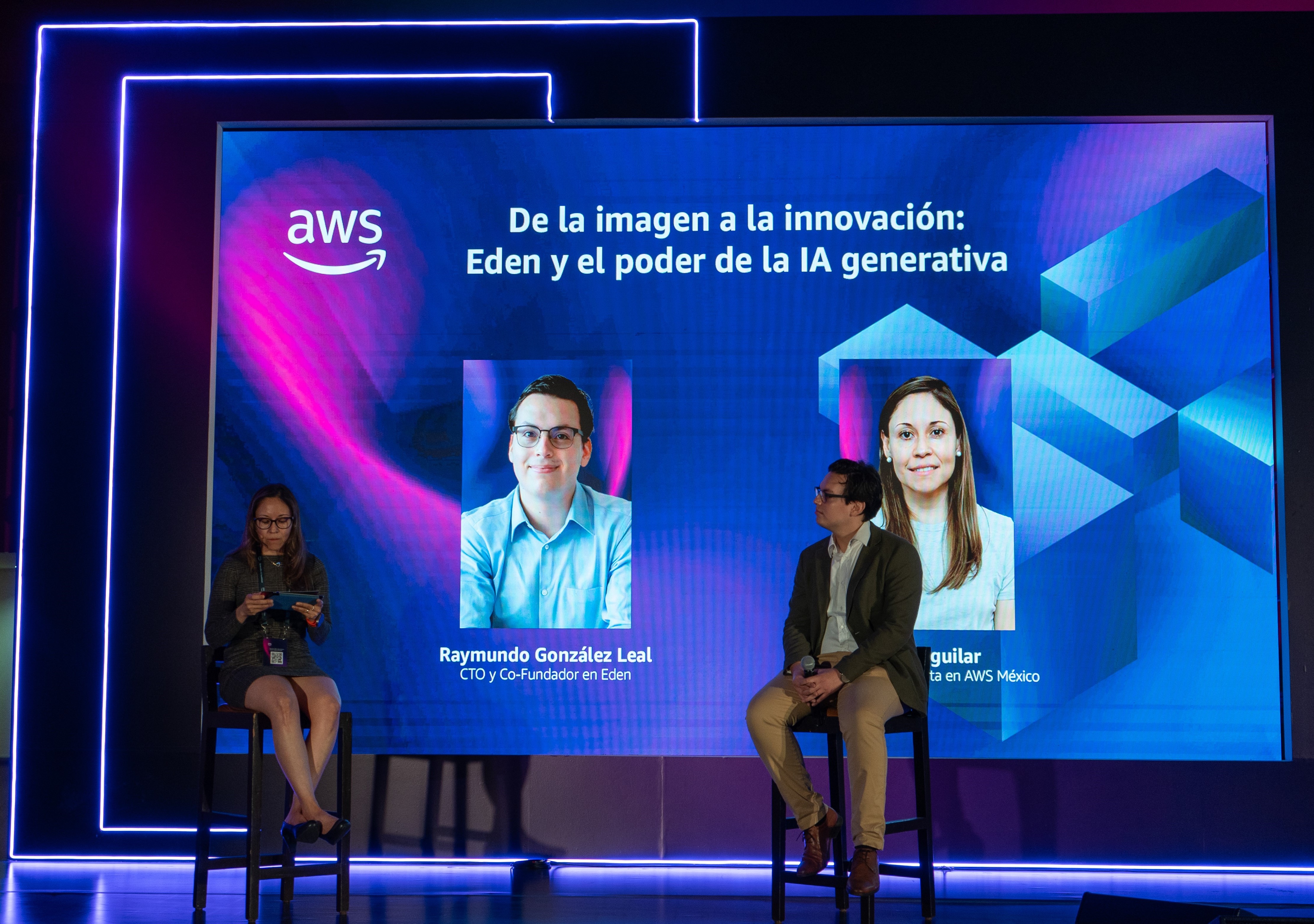
With approximately 4,000 radiologists for 130 million inhabitants, Mexico faces a significant challenge in the healthcare sector. Radiologists spend much of their time on repetitive tasks, which leads to high levels of burnout, long wait times, and elevated costs in the private sector.
In his panel titled “From Imaging to Innovation: Eden and the Power of Generative AI”, Raymundo highlighted the need to integrate new technologies into the daily processes of radiologists across Latin America in order to transform the region's healthcare landscape.
“We can't multiply the number of radiologists by ten, but we can develop technology that increases their productivity and, therefore, their impact on the health of millions.”
Eden is revolutionizing digital healthcare by harnessing the power of the cloud and AI applied to radiology. By streamlining administrative processes and offering cloud-based digital results delivery, our partners have multiplied their care capacity and drastically reduced result turnaround times.
In his conversation with Aida Aguilar, AI sales specialist for AWS, Raymundo also stressed the importance of creating advanced tools that serve radiologists, rather than working against them.
“Many smart solutions tend to be isolated products, adding steps to the physician's workflow instead of reducing their workload.”
With Eden Creator, the generative AI that complements clinical reports with precise conclusions, it is possible to integrate advanced technology naturally into the workflow within a digital ecosystem. In this way, the radiologist saves up to 1 hour of work per day, minimizing the time spent on repetitive tasks and creating new opportunities to improve the quality of patient care.
“It is crucial to give healthcare institutions visibility and transparency in the use of these technologies, provide effective support during implementation, and ensure they can measure whether their objectives are being met.”
At Eden, Artificial Intelligence is more than a productivity tool; it is a definitive step toward a more efficient, effective, and patient-centered healthcare system. We will continue developing innovative tools that improve medical diagnosis, place the patient at the center of care, and respond to the sector's true needs, in order to impact the health of millions.

How to Build Patient Loyalty?
Studies reveal that, for 60% of patients, the way they receive their results is crucial in deciding whether to return to a healthcare institution. Patients want an extraordinary experience that not only delivers their results, but also gives them the power to make informed decisions about their health, all in the palm of their hand.
Accessible
The results delivery experience must be designed with the patient at the center, ensuring an accessible and practical experience. Nearly 80% of patients who decide to switch healthcare institutions have had difficulties using digital platforms or interacting with front-desk staff.
For this reason, to promote loyalty, Eden introduces the most intuitive portal on the market: Eden Portal. This patient portal allows effortless navigation through clinical records and access to results from any mobile device. This streamlines the healthcare experience and boosts satisfaction, encouraging patients to return.
Advanced
It is essential to take advantage of new advanced technologies to improve referral rates and strengthen a medical institution's reputation. It has been shown that when an institution offers an above-average healthcare experience, it can increase its margins by up to 50%1 through greater loyalty, satisfaction, and referral rates.
Eden Portal not only provides this experience to patients, but also to referring physicians. With the world's first DICOM viewer designed exclusively for mobile devices, it is possible to view radiology results instantly and access tools such as 3D reconstruction with a single tap. This allows physicians to do their work more easily and efficiently, maintaining their loyalty and increasing the number of referred patients.
In the Patient's Hands
The true key to a patient-centered experience is giving patients back the power over their own health. This not only increases retention, but also raises the quality of medical care, encouraging patients to get timely exams and stay on top of their well-being.2
With Eden Portal, that power is in your patients' hands, giving them direct, instant access to their entire medical record. This redefines their healthcare experience, making it more personalized, transparent, and centralized.
When an institution places the patient at the center of its operations, the patient will position that institution as the central hub of their healthcare. Eden Portal is designed to be a strategic ally in retention, offering an exceptional experience that gives agency back to the patient. This fosters loyalty and provides patients with the tools they need to take care of themselves, all in the palm of their hand.
References
1Accenture. (2024, April 30). Health experience: The difference between loyalty & leaving | Accenture. Www.accenture.com. https://www.accenture.com/us-en/insights/health/difference-between-loyalty-leaving
2Jha, D., Frye, A. K., & Schlimgen, J. (2017). Evaluating variables of patient experience and the correlation with design. Patient Experience Journal, 4(1), 33–45. https://doi.org/10.35680/2372-0247.1176
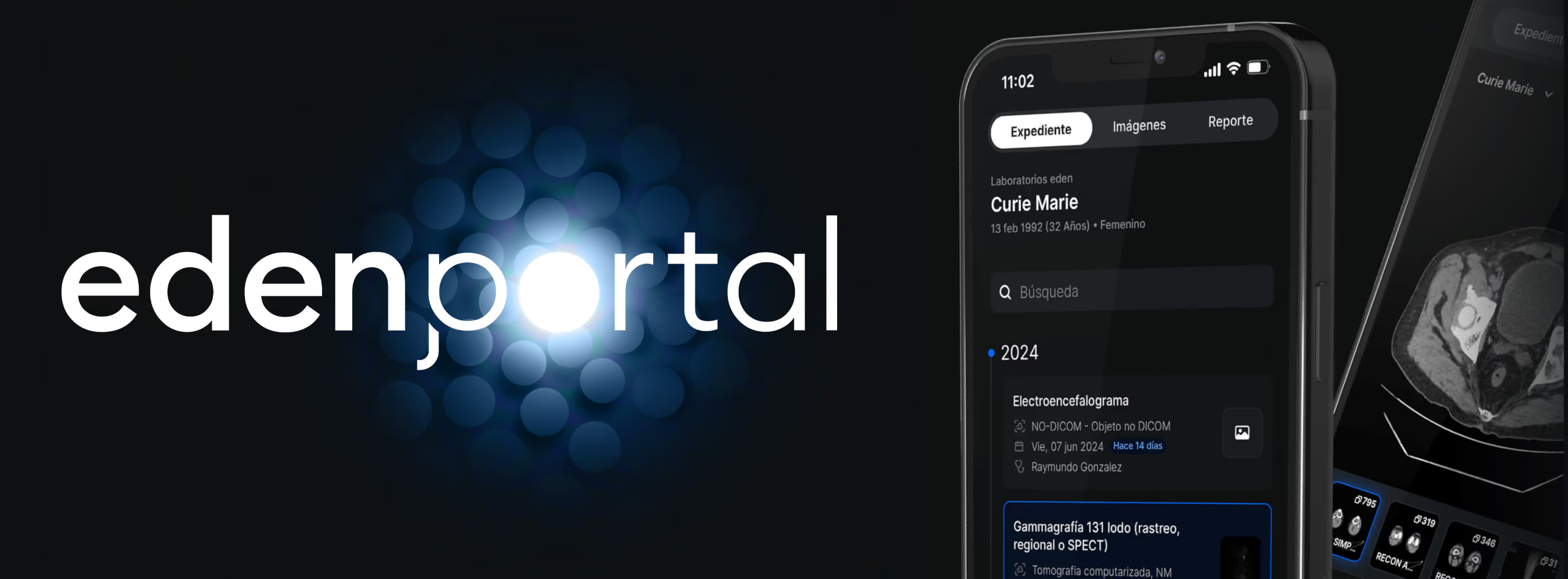
Eden Portal: The Most Powerful Patient Portal on the Market Has Arrived
Most medical interactions begin with an image. According to studies, 60% of patients decide whether they will return to a healthcare institution based on how they receive their results. However, current medical portals are complex and difficult to use, putting patients' health at risk by making it harder to access their information.
Now, that has changed. The days when medical portals could overlook the patient are a thing of the past: Eden Portal has arrived to completely transform the healthcare experience across LATAM.
Give your patients back the power over their health
Eden Portal is designed to give patients back the power over their health. Its intuitive platform makes it easy to navigate between medical reports, radiological images, and their complete medical record. This way, patients don't have to struggle to make sense of their medical information: it's all right in the palm of their hand.
Eden Portal isn't just the most intuitive results experience: it's also the most advanced. With the first DICOM viewer designed 100% for mobile devices, Eden Portal allows hundreds of high-resolution images to be viewed in seconds, even from a smartphone on 3G. This completely redefines the healthcare process, putting the tools patients need to understand and take care of their well-being within reach.
Clinical records anywhere, anytime
With Eden Portal, patients no longer have to search through hundreds of misplaced documents every time they see a new doctor. Now, they can instantly access their entire radiology and medical imaging history, anytime, anywhere.
The system automatically organizes the history by referring physician, study type, and chronology, with clear time indicators to remind patients when the next medical study is due. This way, their healthcare experience is centralized around a single platform and a single institution, radically increasing retention rates.
Advanced tools within reach of a single tap
Eden Portal provides access to tools never before seen in a mobile viewer. From specialized measurements to curved multiplanar reconstruction, referring physicians and even patients will be able to analyze radiological images with just a tap on their mobile device screen.
This strengthens loyalty among treating physicians, who will now have the full power of a medical-grade viewer in their hands. This makes it possible to deliver personalized, agile medical care instantly.
For patients, Eden Portal puts all their radiology and imaging information in the palm of their hand. For healthcare institutions, it represents an unprecedented tool for building loyalty among patients and treating physicians.
Move toward the future. Eden Portal is now available: sign up at edenmed.com/portal or reach out to one of our digital transformation experts to get it. Redefine your standard of results and give your patients an extraordinary experience.
Things That May Be Hindering Your Radiologists' Productivity
Advanced technology does not have to be complex. If your PACS system is causing more problems than benefits for your radiologists, it may be time to evaluate its usability: in other words, how user-friendly it is in day-to-day operations.
Approximately 48% of radiologists are dissatisfied with their RIS/PACS due to its lack of usability. Many systems tend to be slow, prone to technical failures or difficult to navigate, thereby reducing productivity rather than boosting it. To avoid these obstacles, it is necessary to evaluate a PACS's practical usability in the radiologist's daily work.
In terms of a PACS system's diagnostic features, there are several usability indicators that can be useful:
Interpretation time per radiologist
Interpretation time per radiologist, a metric visible in Eden Intelligence, is the leading indicator of agility for determining whether there are areas of opportunity in your institution's operations. Using this measure, it is possible to compare the productivity of different radiologists across study modalities to determine whether longer interpretation times are due to the complexity of a study or to a platform's usability.
Image loading speed and tool usage time
These measures make it possible to know whether the radiologist takes longer than necessary waiting for an image to fully load or searching for the right tool. This indicates a system that is not intuitive, forcing the user to fight against it to do their job and losing precious seconds that could save lives. At the institutional level, these seconds also add up to hours of slow work that not only reduce productivity, but also raise the risk of burnout, eye strain and medical error.
Eden PACS is a platform designed around the user, which is why it includes elements such as a right-click submenu, fast image scrolling and a high-speed DICOM viewer. These tools make interacting with the platform easier, thereby increasing usability and enhancing the radiologist's work.
User satisfaction level
A PACS exists to serve the radiologist: for this reason, their experience is the best indicator of how effective a system truly is. Conducting satisfaction surveys is an important part of implementing medical technology, even when there is no visible reduction in productivity. It is through the radiologist's feedback that we can identify not only how "usable" the platform is on a technical level, but also how it is received among users, referring physicians and patients, what role it plays in daily operations, and how it can help an institution become exceptional.
To successfully implement advanced digital radiology technologies, it is essential to prioritize the human experience at their center. In this way, institutions can boost their productivity and offer humanized, personalized and precise healthcare. Eden PACS's usability goes beyond the number of clicks it takes to complete an interpretation: it is a sign of technology that exists to make the extraordinary accessible, for physicians and patients alike.
References
Bramson, R. T., & Bramson, R. A. (2005). Overcoming Obstacles to Work-Changing Technology Such As PACS and Voice Recognition. American Journal of Roentgenology, 184(6), 1727–1730. https://doi.org/10.2214/ajr.184.6.01841727
Halsted, M. J., & Froehle, C. M. (2008). Design, Implementation, and Assessment of a Radiology Workflow Management System. American Journal of Roentgenology, 191(2), 321–327. https://doi.org/10.2214/ajr.07.3122
Nance Jr, J. W., Meenan, C., & Nagy, P. G. (2013). The Future of the Radiology Information System. American Journal of Roentgenology, 200(5), 1064–1070. https://doi.org/10.2214/ajr.12.10326
.avif)
The Cyberattack Crisis in the Healthcare System: Is It the Physicians' Responsibility?
In 2023, more than 133 million medical records were leaked in the United States alone, exceeding the previous year's figure by 156%. It is time to strengthen cybersecurity in the healthcare system, but what role do radiology institutions play in this monumental task?
Every data breach in the medical industry represents a massive loss of resources, puts millions of patients at risk, and has even resulted in the closure of several medical institutions. Solving this problem once and for all requires global collaboration across the different levels of the healthcare system. However, there are also concrete actions that institutions and individuals can take to defend private information against malicious attacks.
Receiving and providing cybersecurity education
The future of radiology is digital. It is therefore the radiologist's responsibility not only to stay current in their area of specialty, but also to become educated in the use of technology platforms. Cyberattacks can occur in various forms, from phishing to ransomware. Physicians must be able to identify and respond appropriately to warning signs of these potential attacks, such as suspicious emails or unusual behavior in computer systems.
It is also crucial to instill this cybersecurity awareness in patients and referring physicians alike. Particularly when using a PACS system to store and share information, radiologists and institutions alike must foster a culture of cybersecurity. This also involves choosing accessible digital platforms that can be easily used even by the most vulnerable groups, such as elderly patients.
Demanding secure technology and regulatory compliance
Healthcare systems and medical institutions also have a responsibility to implement adequate security measures and provide the infrastructure necessary to effectively protect medical information. This includes investing in advanced security technologies that offer continuous updates and dedicated support for identifying potential cyber risks.
It is especially important to require institutions to work with platforms that meet rigorous cybersecurity standards. Eden PACS complies with both HIPAA regulations and the ISO 27001 standard for information protection systems, meaning its digital defense standards are valid at an international level.
Ultimately, the fight against cyberattacks in the healthcare system is a collaborative effort that requires the active participation of physicians, medical institutions and technology providers. By working together to strengthen cyber defenses and promote a culture of cybersecurity in the field of medicine, it becomes possible to mitigate the risk of data breaches and protect the safety of millions of patients.
References
HIPAA. (2024). SECURITY BREACHES With The Compliments Of The HIPAA Journal IN HEALTHCARE IN 2023. https://www.hipaajournal.com/wp-content/uploads/2024/01/Security_Breaches_In_Healthcare_in_2023_by_The_HIPAA_Journal.pdf
Kesang Tashi Ukyab, Beato, F., & World Economic Forum. (2024, February). Why the healthcare industry must prioritize cyber resilience. World Economic Forum. https://www.weforum.org/agenda/2024/02/healthcare-pays-the-highest-price-of-any-sector-for-cyberattacks-that-why-cyber-resilience-is-key/
Lohchab, H. (2023, November 3). Cyberattacks on healthcare sector rising, 60% of organisations hit in a year: report. The Economic Times. https://economictimes.indiatimes.com/tech/technology/cyberattacks-on-healthcare-sector-rising-60-of-organisations-hit-in-a-year-report/articleshow/104917689.cms?from=mdr
How to Choose the Right PACS for Your Institution's Needs
PACS systems are a fundamental pillar in advancing radiology toward the digital era. However, medical institutions must strategically choose platforms that fit their operational needs. Otherwise, they risk adopting a solution that hinders their productivity rather than boosting it.
It has been shown that the wrong platform can reduce radiologist productivity. Complex navigation, a lack of advanced tools and inadequate support are just some of the shortcomings medical institutions face when implementing this new technology. For this reason, it is necessary to analyze the qualities of a PACS system to explore its maximum potential at each institution.
Interoperability and compatibility
One of the most common problems in implementing PACS systems is viewing the system as an isolated acquisition. In reality, these platforms are part of a whole, both at the technological and operational level. This is why a system's interoperability must be ensured with each institution's radiological equipment, as well as with other everyday platforms used for report dictation, information storage, scheduling and more.
Compatibility must also be considered in terms of who will be using the system and for what purpose. Not all PACS systems have the diagnostic tools suited to a highly specialized radiology unit. Likewise, a PACS with complex navigation can waste crucial seconds for an institution performing emergency radiology. Usability, available tools and even the required level of training are relevant factors that determine a system's long-term accessibility.
Security and regulatory compliance
In a digital medical system, data security must be as robust as the platform itself. It is essential to ensure that a PACS meets basic information protection standards, including data encryption and user authentication. In addition, compliance with privacy regulations such as HIPAA is indispensable. Eden PACS is certified under these regulations as well as the ISO 27001 standard for cybersecurity systems, thereby guaranteeing the privacy of patients and institutions at an international level.
Technical support and customer service
The availability of technical support is one of the most important factors in the acceptance and continued use of new technologies. Eden PACS's 24/7 support resolves technical issues within moments, thereby minimizing downtime and preventing interruptions in patient care. In addition, with ongoing support from expert engineers, system users are able to learn and make the most of all of the PACS's features. For this reason, the quality of technical support affects not only operational efficiency, but also the quality of patient care and the satisfaction of medical staff.
As the range of medical technology available continues to grow, it becomes increasingly important to determine which PACS is right for enhancing your operations. Above all, any system implemented must be adaptable, dynamic and responsive to your institution's needs. A platform that evolves alongside your physicians and users is one that can propel your practice into the future, rather than keeping it anchored in the past.
References
Cox, B., & Dawe, N. (2002). Evaluation of the impact of a PACS system on an intensive care unit. Journal of Management in Medicine, 16(2/3), 199–205. https://doi.org/10.1108/02689230210434934
Duyck, P., Pynoo, B., Devolder, P., Voet, T., Adang, L., Ovaere, D., & Vercruysse, J. (2008). Monitoring the PACS Implementation Process in a Large University Hospital—Discrepancies Between Radiologists and Physicians. Journal of Digital Imaging, 23(1), 73–80. https://doi.org/10.1007/s10278-008-9163-7
.avif)
How to Get the Most Out of the 3D Reconstruction Tool
Volumetric reconstruction is a powerful tool that, now available in the Eden PACS DICOM viewer, is within reach of more physicians than ever before. This opens up new opportunities to integrate 3D visualizations into radiological practice and raise the standard of care at institutions throughout Latin America.
3D reconstruction has primarily been used to achieve precise interpretations in fields such as cardiology and orthopedics. However, it also offers opportunities for growth in two vital areas beyond diagnosis: connecting with specialist physicians and communicating with patients.
Connections for personalized medical care
3D visualizations are invaluable in preoperative planning and interventional radiology procedures. A three-dimensional image makes it easier to identify critical structures in a patient's anatomy and even allows different surgical approaches to be simulated, transforming the precision and safety of these treatments.
Radiology clinics can use these capabilities as a connection point with other institutions and specialist physicians. This would not only raise their revenue, influence and impact, but would also represent a turning point for personalized medicine in Latin America. The 3D tool makes it possible to account for each patient's unique anatomy and facilitates communication among their physicians. In this way, it becomes possible to provide precise follow-up and improve the health of thousands of patients.
Putting the patient at the center of care
Recent studies on 3D reconstruction emphasize its usefulness in explaining medical conditions to patients. By showing them a three-dimensional representation of their anatomy, it becomes possible to help them better understand their diagnosis and recommended treatment plan.
This level of communication leads to better clinical outcomes, motivates patients to follow instructions and raises their satisfaction levels. It is also an approach that helps transform current healthcare processes, as it invites patients to become part of their own care and to feel empowered within a system that is often dehumanizing.
Radiological technology is not reserved for the reading room. Tools such as 3D reconstruction can revolutionize the way healthcare is delivered and received. By harnessing its potential across different areas of the system, it becomes possible to bring exceptional healthcare to institutions throughout the continent.
References
Haleem, A., Javaid, M., Suman, R., & Singh, R. P. (2021). 3D Printing Applications for Radiology: An Overview. The Indian Journal of Radiology & Imaging, 31(1), 10–17. https://doi.org/10.1055/s-0041-1729129
Jones, J. (2022, March). Creating 3D Models from Imaging. Www.acr.org. https://www.acr.org/Practice-Management-Quality-Informatics/Imaging-3/Case-Studies/Information-Technology/Creating-3D-Models-from-Imaging
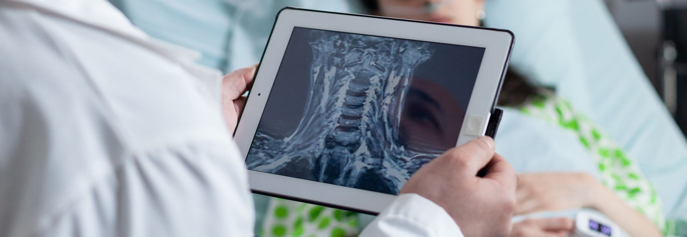
The Imminent Changes the Medical System Needs to Provide Access to Radiology
Access to quality radiology remains a dream for much of Latin America.
It is estimated that conditions whose diagnosis relies on imaging studies, such as cancer and cardiovascular disease, are responsible for nearly 82% of deaths in Latin America and the Caribbean. In Mexico alone, there are 30 radiologists and three magnetic resonance imaging (MRI) units per million inhabitants. Across the continent, medical systems have become increasingly costly, saturated and limited in their capacity to provide care.
Radiology is regarded by many as the medicine of the future because of its continuous technological advances. But if basic care remains out of reach for the general population in the developing world, this evolution will be in vain. To truly bring the future to everyone, it is necessary to transform the medical system and restructure the way technology is selected, adopted and implemented according to the region's unique needs.
Medicine at the service of the patient
In an overwhelmed system, patients are no longer at the center of medical care. Many must wait months to undergo radiological exams, incur significant costs traveling to healthcare centers, and frequently receive delayed, incorrect or incomplete diagnoses.
Addressing these problems requires adopting technologies aimed at reducing administrative burden and improving the patient experience. In the case of Eden Management, the platform is designed to centralize operations with minimal infrastructure, allowing implementation in any corner of the continent. This, combined with the digital delivery of results via WhatsApp, is a solution that simplifies the patient experience within the healthcare system's workflow and speeds up the diagnostic process for timely medical care.
Tools at the service of the physician
A central element in the adoption of new technologies is the radiologist's experience. In Latin America, radiologists are overburdened with work and many lack the equipment needed to perform it properly. As a result, costly, complex technologies with high infrastructure requirements would become an obstacle rather than a productivity tool.
For this reason, Eden PACS was created to be intuitive, lightweight and accessible from any device. These strategies help work around infrastructure challenges to offer physicians intuitive diagnostic tools. Likewise, with the implementation of artificial intelligence in the form of Eden Creator, it is possible to reduce workload and take the first step toward automating repetitive processes.
Technology at the service of everyone
The vast majority of advanced radiological technology providers are large international companies whose products are not adapted to the needs of the developing world. The giants of the medical industry see only what is in their line of sight.
That is why Eden was born: to cover the medical system's blind spots in Latin America from within. By building a close relationship with our partner institutions, we can respond directly to the experiences of physicians and patients throughout the region. It is only through this continuous evolution, adaptability and closeness to the realities of the continent that it will be possible to bring extraordinary radiology to everyone.
References
McKenney, A. S., Garg, T., Kim, E., & Kesselman, A. (2021). Addressing Global Radiology Disparities: Increasing Access to Interventional Radiology Education. RadioGraphics, 41(5), E142–E144. https://doi.org/10.1148/rg.2021210176
Murali, S., Ding, H., Adedeji, F. A., Qin, C., Obungoloch, J., Asllani, I., Anazodo, U., Ntusi, N., Mammen, R., Niendorf, T., & Adeleke, S. (2023). Bringing MRI to low‐ and middle‐income countries: Directions, challenges and potential solutions. NMR in Biomedicine. https://doi.org/10.1002/nbm.4992
Rodriguez, A., Martínez, L., & Alvarado, S. R. (2023). Uso de nuevas tecnologías en Radiología e imágenes diagnósticas y su relación con las competencias profesionales y/o perfil de egreso del Licenciado en Radiología de Panamá y Latinoamérica en los últimos 15 años. Ciencia Latina Revista Científica Multidisciplinar, 7(1), 6762–6788. https://doi.org/10.37811/cl_rcm.v7i1.4929
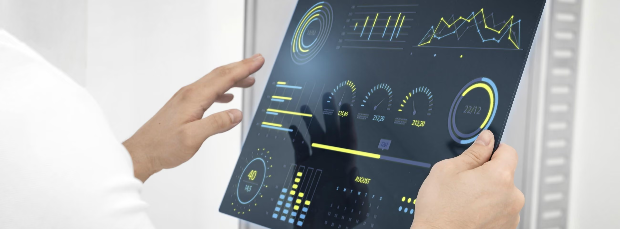
How Does Business Intelligence Elevate Radiology?
Business intelligence is more than a tool for making business decisions. Especially in the field of radiology, it has become clear that this data must be used in strategies that go beyond productivity and aim to elevate the quality of medical work. With the right metrics, this technology can reveal critical factors in the radiological process, helping create more value for both physicians and patients.
What is business intelligence?
Business intelligence, or BI, refers to a set of technologies and tools that help organizations collect, analyze, and present data to make informed decisions. However, BI solutions specialized for radiology, such as Eden Intelligence, focus on measuring precise indicators for the industry, including the number of studies by modality and the number of studies interpreted per physician, among others.
Metrics that go beyond productivity
This data is necessary to increase productivity, reduce operating costs, and evaluate the performance of multiple branches, especially in the case of clinical laboratories. However, its specificity also makes it possible to assess how these institutions operate around their patients’ needs. Based on these metrics, radiologists themselves can make adjustments to their way of working and contribute ideas for process improvement based on hard, real-time data. For example:
- The individual productivity of each member of the medical staff reveals not only performance quality, but also workload distribution and demand for certain specialists
- The number of studies performed per hour helps identify bottlenecks and assess the effectiveness of work plans, shift assignments, and staff rotation
- Trends in the modality of studies performed provide general indicators of current demand and help assess the need for different pieces of medical equipment and/or personnel
In addition to these metrics, Eden Intelligence includes two key indicators: physician interpretation time and number of referred studies. From these, factors in customer satisfaction can be extrapolated, since patients depend on the physician’s interpretation time to receive their studies. Likewise, the number of referred studies is a criterion for evaluating an institution’s relationship with external physicians: their trust in the radiologist, the loyalty established with the institution, and their satisfaction with its digital referral system.
The future of BI in radiology
These elements transform BI for radiologists, as they go beyond organizational boundaries to explore the satisfaction of partners and patients. In the future, platforms like Eden Intelligence will continue to evolve to include specific metrics that make it possible to interpret medical work not only at the business level, but also in terms of the value it delivers. Only in this way will healthcare institutions be able to move beyond good and into the extraordinary.
References
Boland, G. W., Thrall, J. H., & Duszak, R. Jr. (2014). Business Intelligence, Data Mining, and Future Trends. American College of Radiology. https://www.acr.org/Practice-Management-Quality-Informatics/Imaging-3/Imaging-Value-Chain/Data-Mining
Yee, J. M., Cross, N., & Bhargava, P. (2022). Do-It-Yourself Business Intelligence for the Radiologist—Lessons Learned From 10-Year Trends in an Abdominal Imaging Division at a Tertiary Medical Center. Journal of the American College of Radiology, 19(2), 329–335. https://doi.org/10.1016/j.jacr.2021.10.007
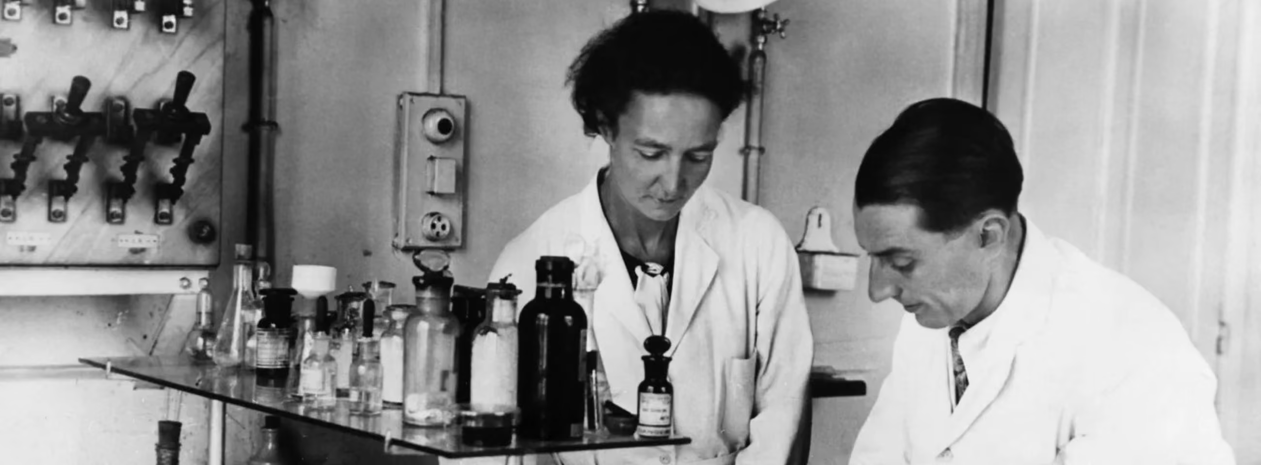
Pioneers in Radiology: 5 Women Who Changed Everything
On International Women's Day, we honor the achievements of five women who transformed radiology. Thanks to their discoveries, contributions, and inventions, millions of patients can access diagnostic imaging and radiotherapy services. Let us continue working to increase this number and to honor the great physicians and scientists who elevated radiology to new heights.
Irène Joliot-Curie, D.Sc (1897-1956)
Dr. Joliot-Curie’s achievements speak for themselves. In addition to serving as a field radiologist during World War I, she carried on her mother Marie Curie’s legacy in the study of radioactivity. Together with her husband Frédéric, she discovered the existence of radioactive isotopes that could be manufactured at low cost. This breakthrough made imaging and cancer treatments accessible for the first time, earning her the Nobel Prize in 1935. During the ceremony, the committee declared that Dr. Joliot-Curie’s discoveries would lead to “remedies of inestimable value” for all of humanity, and so they did.
Edith Quimby, D.Sc (1891-1982)
Dr. Quimby’s specialty was physics, but her domain was medicine. Her work grew out of a desire to protect both physicians and patients from the harm of X-ray radiation without compromising the quality of treatment. Quimby’s research transformed the landscape of dosimetry and forms the basis of the Memorial system, still used today to calculate radiation doses in brachytherapy. Thanks to her, diagnostic radiology exists as we know it today: effective, advanced, and above all, safe.
Tikvah Alper, M.A. (1909-1995)
Dr. Alper made the invisible visible through radiobiology. During her research on the effects of radiation on cells, she discovered that certain viruses can replicate without being affected by radiation. This revolutionized the field and paved the way for prion theory, which today forms the basis of our understanding of infectious neurodegenerative diseases. Alper was not afraid to question established knowledge and go further to uncover the truth, breaking paradigms and taking radiobiology to another level.
Alice Ettinger, MD (1899-1993)
Dr. Ettinger was a hero in more ways than one. She arrived in the United States from Berlin on what she thought would be a trip of a few weeks, and ended up staying for more than 50 years at Tufts University School of Medicine. She introduced gastrointestinal radiology to the country, pioneered the emerging field of tomography, and gave work to doctors who had fled Germany during the 1930s. Even today, Ettinger has left her mark among colleagues as an extraordinary radiologist who propelled the discipline to new horizons.
Muyinatu Bell, PhD
Dr. Bell is responsible for the most transformative innovations in radiological equipment in recent years. She is the founder and director of the Photoacoustic and Ultrasonic Systems Engineering (PULSE) Laboratory at Johns Hopkins University. She also led the development of the first Short-Lag Spatial Coherence (SLSC) system for ultrasound, introduced the use of fiber-optic photoacoustic imaging to assist in delicate surgeries, and is a pioneer in combining these tools with minimally invasive robotic surgery. Still active as a researcher, Bell promises to be an essential force in creating new advanced radiology tools.
References
Coultas, P. G., & Hopewell, J. W. (2021). Dr Tikvah Alper: a short history of her scientific career. International Journal of Radiation Biology, 98(3), 314–317. https://doi.org/10.1080/09553002.2021.2009148
Johns Hopkins Whiting School of Engineering. (2024). Muyinatu Bell. Johns Hopkins Whiting School of Engineering. https://engineering.jhu.edu/faculty/muyinatu-bell/
Karakatsanis, N. A., & Arleo, E. K. (2022). Dr. Edith H. Quimby: A pioneering medical physicist and educator with outstanding contributions in radiation dosimetry. Clinical Imaging, 81, 118–121. https://doi.org/10.1016/j.clinimag.2021.09.017
Science History Institute. (n.d.). Irène Joliot-Curie and Frédéric Joliot. Science History Institute. https://www.sciencehistory.org/education/scientific-biographies/irene-joliot-curie-and-frederic-joliot/#:~:text=Partners%20in%20life%20and%20in
U.S. National Library of Medicine. (2015a). Alice Ettinger. Changing the Face of Medicine. https://cfmedicine.nlm.nih.gov/physicians/biography_105.html
No papers match your search.